Synthesis and X-ray structure
of 3, 3'-diethoxycarbonyl-2, 2'- diethoxy-1, 1'- binaphthyl
Li Lijuna, Li Cuizhea, Zhang Dahaib,
Zang Jiechaoa
(a, College of Chemistry and Environment Science, Hebei University, Baoding 071002; b,
department of chemistry, Handan college, Handan 056005)
Received Nov. 28, 2008; Supported by the fund
from the Natural Science Foundation of Hebei Province (No. B2007000152) and Plan Item of
Hebei Province Science-Technology Department (05215104)
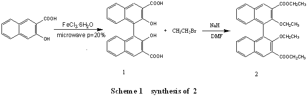
Abstract A novel 3,3'-BINOL derivative:
3,3'-diethoxycarbonyl-2,2'- diethoxy-1,1'- binaphthyl(2) was synthesized and its structure
was characterized by IR, 1H NMR, 13C NMR and X-ray diffraction.
Keyword 3, 3'-diethoxycarbonyl-2, 2'- diethoxy-1, 1'- binaphthyl, synthesis,
characterization
1 INTRODUCTION
Optically active BINOL and its derivatives are the most prominent C2-symmetric,
axial-chiral ligands,they are
well known to apply in asymmetric catalysis and chiral recognition[1-4]
. This
is because: BINOL and its derivatives have a chair backbone which can be modified.
Substitution of BINOL may affect not only the steric environment around the coordinated
metal center but also the electronic properties of the oxygen atoms, which are common
constituents of the Lewis acidic metal complexes [5-6]. BINOL itself, however,
does not always give satisfactory results in asymmetric catalysis, and there has been an
ongoing interest in modified BINOL ligands[7-8] since people's discovery.
This paper firstly
synthesized BINOL-3,3'-dicarboxylic acid (1) under microwave irradiation with
iron(III) chloride hexahydrate as catalyst, then a new BINOL derivative
3,3'-diethoxycarbonyl-2,2'- diethoxy-1,1'- binaphthyl (2) was synthesized by substitution
reaction.The synthetic routes were outlined in Scheme 1. And its structure was characterized through IR, 1H
NMR, 13C NMR and X-ray diffraction, and emphatically analyses its X-ray date.
2. EXPERIMENT
2.1 Materials and general methods
Commercial reagents were used without further purification unless otherwise noted. The
melting points were determined using a digital melting point apparatus. IR spectrawere
recorded on a Nicolet AVATAR-37ODTGS Spectrophotometer in the region 4000-400 cm-1 KBr discs. 1H NMR and 13C
NMR spectra were recorded on a Bruker Avance-400 superconduct nuclear magnetic resonance
instrument, in which chemical shift values are reported in ppm (d) relative to TMS as
internal standard. X-ray date was recorded on Rigaku Saturn X ray diffractometer at room
temperature.
2.2 Syntheses of 1
The BINOL-3, 3'-dicarboxylic acid was synthsized
according to reported mothed [9]. Iron(
III) chloride hexahydrate (20.3g, 56mmol) and 3- hydroxy-2- naphthoic acid fully mixed grind, the mixture was
transferred to 100mL beaker, put into a commercial microwave oven Galanz WD800G, set
P=20%, t=30s, took out the breaker and stired fully, then put into microwave oven,
repeatedly ,till the time of reaction arrived to 740s. The solid was washed by 100mL mixed
solution of 36%HCl and H2O (v/v=1/1), and was collected by vacuum filtration,
then purified by column chromatography using ethyl acetate: petroleum ether (3:1, v/v) as
the eluent. The main peak was concentrated to afford BINOL-3, 3'-dicarboxylic acid (7.0 g, 18.7mmol) as a yellow solid. Yield: 91%;
Mp 295-297oC (Lit.[10]Mp>290oC); 1H NMR
((CD3)2CO, 400 MHz): d 7. 14-7. 17
(m ,2H ,Ar-H) ,7. 39-7. 42(m ,4H ,Ar-H) ,8.
09-8. 11(m ,2H ,Ar-H) ,8. 84( s ,2H ,Ar-H);
IR ( KBr) 3059( s ,nO-H) ,1661( s ,nC =O) ,1499,1455 , 1 272, 1 228 , 1 150 , 1 072
, 886 , 796 , 736cm- 1 .
2.3 Syntheses of 2
At room temperature, compound 1 (0.37g, 1mmol) and NaH(52% dispersion in mineral oil;
0.12g, 2.62 mmol) was added in 5mLDMF, then the mixture was allowed to warm to 50oC, and
stirring for 1.5 h.. After that 1.5mL CH3CH2Br was slowly added.
Every hour NaH 80mg (1.78mmol) was added in mixture. Tracking the reaction by TLC, until
compound 1 reacted completely. Then 10mL H2O was added to the mixture,
extracted with ethyl acetate (3×50mL). The combined organic layers were dried over MgSO4
and the solvent was removed under reduced pressure. The solid residue was dissolved in
ethyl acetate and purified by column chromatography to afford the compound 2 (0.37g, 77%)
as a white solid; Mp:104oC.
IR (KBr):2978, 2932, 2898,1725, 1622,1587,1447,1385, 1364, 1339, 1296, 1271, 1227, 1145,
1108, 1070, 1033, 907, 795, 754, 711cm-1; 1H NMR(CDCl3 ,
500MHz) d: 0.78-0.81 (t ,3H ,COOCH2CH3) ,1.42-1.45 (t,
3H, OCH2CH3), 3.47-3.53 (m, 2H, OCH2CH3),
3.87-3.93 (m, 2H, OCH2CH3), 4.43-4.47 (q, 4H, COOCH2CH3
), 7.13-7.15 (d ,2H ,Ar-H) ,7. 30~7.33(m ,2H ,Ar-H)
, 7.41-7,44 (m ,2H ,Ar-H), 7. 95-7. 96(d , 2H ,Ar-H) , 8. 48 ( s , 2H , Ar-H); 13C NMR
(CDCl3,125 MHz) d: 14.3, 15.3, 61.3, 70.3, 77.0, 125.3, 125.7, 125.9, 126.5,
128.2, 129.0, 129.4, 132.7, 135.6,153.7, 166.8.
2.4 X-ray crystallography
Crystals of compounds 2 were grown by liquid–liquid diffusion. Mixed solvent of DMSO:H2O=1:1(v/v)
Table 1. Crystal data and structure
refinement for 2
Empirical formula |
C30H30O6 |
q range for data collection |
2.71 to 27.63° |
Formula weight |
486.54 |
Limiting indices |
-14 < h < 12 |
Temperature (K) |
293(2) |
|
-20 < k < 20 |
Wavelength(Å) |
0.71073 |
|
-20 < l < 20 |
Crystal system |
Monoclinic |
Reflections collected |
19223 |
Space group |
P21 /n |
Independent reflections |
5903 (Rint = 0.0397) |
a( Å) |
11.138(18) |
Completeness to q |
97.8 % |
b( Å) |
15.60(3) |
Goodness-of-fit on F2 |
1.061 |
c( Å) |
15.80(3) |
Data / restraints / parameters |
5903 / 0 / 330 |
Volume( Å3) |
2596(7) |
R1(R1 all data) |
0.0915 |
Z |
4 |
wR2 |
0.1980 |
Dcalcd (Mg/m3) |
1.254 |
Largest diff. peak and
hole eÅ-3 |
0.267 and -0.241 |
m (mm-1) |
0.086 |
F(000) |
1032 |
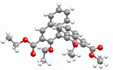
Table2 Selected bond and distance (
Å) and angles (°) of 2
C(1)-C(11) |
1.496(4) |
C(21)-O(6) |
1.204(3) |
C(22)-O(1) |
1.189(3) |
O(4)-O(6) |
2.865 |
O(2)-O(3) |
2.730 |
|
|
|
|
|
|
C(2)-C(1)-C(11) |
120.62(19) |
C(10)-C(1)-C(11) |
118.56(16) |
C(20)-C(11)-C(1) |
119.03(06) |
C(12)-C(11)-C(1) |
120.16(17) |
C(12)-O(4)-C(27) |
116.34(17) |
C(2)-O(3)-C(25) |
115.63(17) |
O(5)-C(11)-O(6) |
2.246(4) |
O(1)-C(22)-O(2) |
121.6(2) |
2 mL was added to the tube; compounds2
(9.7mg, 0.02mmol) were dissolved in 2ml DMF, was then slowly added along the sides of the
tube via a pipette so that the solution was not disturbed, forming the upper layer. The
tube was then covered and the solvents were allowed to slowly diffuse at room temperature.
After 7 days, colorless prism crystal was found.
The resultant crystals were separated from the solution by decanting.
Suitable X-ray quality crystals were recovered and mounted using epoxy glue. Data were
collected on a Bruker SMART APEX CCD area detector diffractometer. SAINT (Bruker, 1998),
SHELXTL (Bruker, 1998) and SHELXS 97 [11-12] were used for cell refinement,
data reduction and structure solving and refinement of structure. Molecular graphics and
publication materials were prepared using SHELXTL [13]. Details of the crystal
data are listed in Table 1.Selected bond lengths and angles are listed in Table
2.
3 RESULTS AND DISCUSSION
3.1 Syntheses and spectral analyses
Firstly, we synthesized the compound 1 according to reported mothed [9], when setting the
power of microwave oven with 20%, the mole ratio n(Iron(
III)
chloride hexahydrate): n(3-hydroxy-2-naphthoic acid)=1:1.5, the yield was the highest 74%
after 740s. The yield had been improved compared with Villemin's report, and the product
gives well-resolved 1HNMR spectra consistent with the report of Jingzhong Yi
[10].
In the synthesis of
compounds 2, we chose NaH as strong base which can seize the hydrogen at hydroxyl and
carboxyl, and it was needed to add intermittently. If the NaH was added much more one
time, the DMF would be deduced. If the NaH was added much less, the reaction time would be
longer. The compound 2 structure was characterized by IR, 1H NMR, and 13C
NMR. The BINOL derivative gives well-resolved 1HNMR spectra consistent with its
structure.
3.2 Crystal structures of compound 2
The crystal structure of 2 shows in Figure1,
in which the substitutes of naphthalene ring is far, its configuration is R-2, and the
dihedral angle of two naphthyl rings is 87.44(9)o, nearly perpendicular.
Figure 1
the molecular structure of 2
In the crystal cell of 2, every two
molecules form a small cuboid channel, four molecules form a bigger cuboid channel, and
there are two naphthyl rings are parallel in two adjacent molecules (see Firgure 2
and
Firgure 3).
Figure 2 crystal cell of 2 along a axis
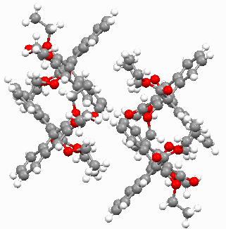
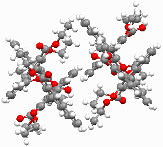
Figure 3 crystal cell of 2
along b axis
REFERENCES
[1] Maki M, Takeshi N. Tetrahedron Lett., 38 (1997): 6233.
[2] Pu L. Chem. Rev., 98 (1998): 2405.
[3] Junji I, Hiroshi F, Tetsuji H. Chem. Rev., 102 (2002): 2211.
[4] Helen C A. Chem. Rev. 102 (2002): 1807.
[5] Haruro I, Yasuhiro Y, Haruka S, etal. J. Am. Chem. Soc., 122 (2000):
5403.
[6] Ana M, Karl H D. Tetrahedron: Asymmetry, 16 (2005): 3256.
[7] Yu C, Shahla Y, Andrei K, etal. Chem. Rev., 103 (2003):3155.
[8 Pavel K, Sÿt
êpán
V, Martin S. Chem. Rev., 103(2003): 3213-3245.
[9] Didier V, Frédérique S. Synlett, 6(1994): 435.
[10] Yi J Z, Liu S, Chen A Q. Jounal of Xiamen University (Natural Sciences), 43(2004):
652.
[11] Sheldrick G.M., SHELXS 97, Program for Crystal Structure Solution. Göttingen
University, 1997.
[12] Sheldrick G.M., SHELXS 97, Program for Crystal Structure Refinement. Göttingen University, 1997.
[13] Sheldrick G.M., SHELXS Version 5, Siemens Industral Automation Inc. Madsion, WI,
1995.
3,3'-二乙氧甲酰基-2,2'- 二羟基-1,1'-联二萘的合成和单晶结构
李立军a
李翠哲a 张大海b 臧杰超a
(a河北大学化学与环境科学学院
保定071002 b 邯郸大学化学系 邯郸056005 )
摘要 合成了一种新的3,3'-取代的BINOL衍生物,3,3'-二乙氧甲酰基-2,2'- 二羟基-1,1'′- 联二萘(2),并且通过IR, 1H NMR, 13C NMR和X-ray单晶衍射等方法对其结构进行了表征
关键词 3,3'-二乙氧甲酰基-2,2'- 二羟基-1,1'- 联二萘;合成;单晶结构